
These observations suggest that contrast enhancement at MR imaging may be influenced by factors that have prognostic value. Furthermore, there was a correlation between contrast enhancement and tumor malignancy grade as well as tumor invasiveness. There was significant correlation between contrast enhancement at MR imaging of breast cancer and both tumor angiogenesis and proliferative cellular activity as shown by PCNA immunoreactivity. We compared contrast enhancement in 50 malignant breast tumors at MR imaging to several prognostic factors, such as tumor size, lymph-node status, histological grade of malignancy, tumor angiogenesis, and proliferating activity as shown by the mitotic count and PCNA immunoreactivity. Thus certain characteristics such as tumor angiogenesis and the proliferating activity of the tumor, which have been shown to correlate significantly with prognosis, are both potentially amenable to analysis by MR imaging. Using contrast-enhanced MR imaging in the diagnosis of breast cancer may provide additional information not only on tumor extension but also on the biological behavior of tumors. This technique allows regional perfusion maps to be measured noninvasively, with the resolution of 1H MRI, and should be readily applicable to human studies. min−1 (mean ± SEM, n = 3), in good agreement with values reported in the literature, and was sensitive to increases in arterial pCO2. Average cerebral blood flow in normocapnic rat brain under halothane anesthesia was determined to be 105 ± 16 cc. Distal saturation applied equidistantly outside the brain serves as a control for effects of the saturation pulses. Because proton T1 times are relatively long, particularly at high field strengths, saturated spins exchange with bulk water in the brain and a steady state is created where the regional concentration of saturated spins is determined by the regional blood flow and regional T1. Blood water flowing to the brain is saturated in the neck region with a sliceselective saturation imaging sequence, creating an endogenous tracer in the form of proximally saturated spins.

Here we report the use of 1H NMR imaging to generate perfusion maps in the rat brain at 4.7 T.
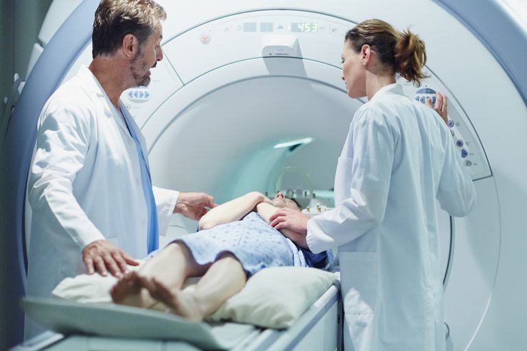
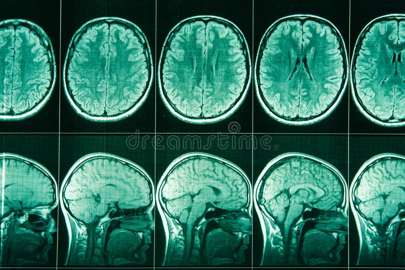
Mri con contraste de la cabeza full#
See /license for the full LOINC copyright and license.Measurement of tissue perfusion is important for the functional assessment of organs in vivo.

and the Logical Observation Identifiers Names and Codes (LOINC) Committee. To the extent included herein, the LOINC table and LOINC codes are copyright © 1995-2022, Regenstrief Institute, Inc. LOINC FHIR ® API Example - CodeSystem Request Get Info $lookup?system= LOINC CopyrightĬopyright © 2022 Regenstrief Institute, Inc. Language Variants Get Info zh-CNChinese (China) 多层^不采用对比剂: 发现: 时间点: 头部>脑部: 文档型: MR it-ITItalian (Italy) Sezioni multiple^Senza contrasto: Osservazione: Pt: Testa>Cervello: Doc: MR pt-BRPortuguese (Brazil) Multi cortes^sem contraste: Achado: Pt: Cérebro: Nar: RM es-ARSpanish (Argentina) ^sin contraste: hallazgo: punto en el tiempo: cerebro: Narrativo: resonancia magnética es-MXSpanish (Mexico) Contraste multisección ^ WO: Tipo: Punto temporal: Cabeza> Cerebro: Documento: MR Related Names Observation Both HL7 Attachment Structure Implementation guide exists Member of these Groups LG41824-0 The LOINC/RadLex Committee agreed to use a subset of the two-letter DICOM modality codes as the primary modality identifier. Changed System from "Brain" to "Head>Brain" for conformance with LOINC/RadLex unified model. LOINCĭiagnostic imaging report - recommended C-CDA R1.1 sectionsīasic Attributes Class RAD Type Clinical First Released Version 2.04 Last Updated Version 2.61 Change Reason The scale has been changed from "Nar" to "Doc" to fit with the CDA model. This panel contains the recommended sections for diagnostic imaging reports based on the HL7 Implementation Guide for CDA® Release 2: Consolidated CDA Templates for Clinical Notes (US Realm) DSTU Release 1.1.

LOINCĭiagnostic imaging report - recommended C-CDA R2.0 and R2.1 sectionsĬurrent imaging procedure descriptions Document This panel contains the recommended sections for diagnostic imaging reports based on HL7 Implementation Guide for CDA® Release 2: Consolidated CDA Templates for Clinical Notes (US Realm) DSTU Releases 2.0 & 2.1. Version 2.72 30657-1 MR Brain WO contrast Active Fully-Specified Name Component Multisection^WO contrast Property Find Time Pt System Head>Brain Scale Doc Method MR Additional Names Short Name MR Brain WO contr Associated Observations


 0 kommentar(er)
